
Image: Cartilage from mouse joint
Size of this preview: 800 × 600 pixels. Other resolutions: 320 × 240 pixels | 2,400 × 1,800 pixels.
Original image (2,400 × 1,800 pixels, file size: 1.06 MB, MIME type: image/jpeg)
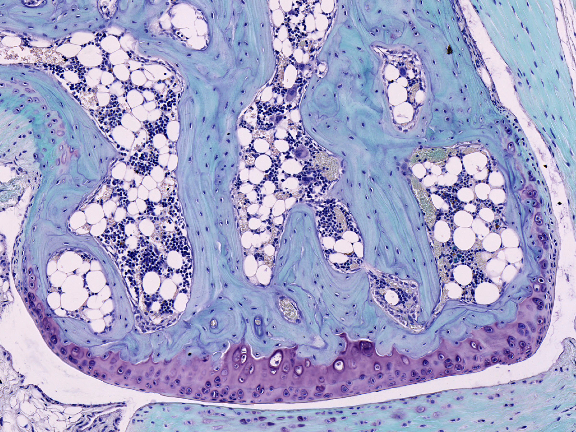
Description: Section through the knee joint from a mouse stained with Safranin O and light green solution. The bone is stained pale blue and the cell nuclei of the bone and bone marrow are visible as small dark blue spots. The cartilage on the edge of the joint (purple) and fat cells (large white spaces) are also visible here. Width of image is 1 mm.
Title: Cartilage from mouse joint
Credit: https://wellcomecollection.org/works/zc8ux23k
Author: Kevin Mackenzie, University of Aberdeen
Usage Terms: Creative Commons Attribution 4.0
License: CC BY 4.0
License Link: https://creativecommons.org/licenses/by/4.0
Attribution Required?: Yes
Image usage
The following page links to this image:

All content from Kiddle encyclopedia articles (including the article images and facts) can be freely used under Attribution-ShareAlike license, unless stated otherwise.