
Image: Chytridiomycosis2
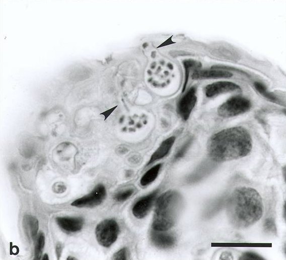
Description: Chytridiomycosis. Ventral skin of upper hind limb of Atelopus varius from western Panama. Two sporangia (spore-containing bodies of Batrachochytrium sp.) containing numerous zoospores are visible within cells of the stratum corneum. Each flask-shaped sporangium has a single characteristic discharge tube (arrow) at the skin surface. Exiting zoospores are visible in the discharge tubes of both sporangia. Hyperkeratosis is minimal in this acute infection. Tissues were fixed in neutral-buffered 10% formalin, paraffin-embedded, sectioned at 6 µm thick and stained with hematoxylin and eosin. Bar = 35 µm.
Title: Chytridiomycosis2
Credit: Daszak P, Berger L, Cunningham A, Hyatt A, Green D, Speare R. (1999). "Emerging Infectious Diseases and Amphibian Population Declines". Emerging Infectious Diseases 5 (6): 735–748. DOI:10.3201/eid0506.990601.
Author: Peter Daszak, Lee Berger, Andrew A. Cunningham, Alex D. Hyatt, D. Earl Green, and Rick Speare
Permission: Public Domain rationale
Usage Terms: Public domain
License: Public domain
Attribution Required?: No
Image usage
The following 2 pages link to this image:
