
Image: Escherichia coli with phages

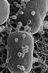
Description: Helium ion microscopy image showing T4 phage infecting Escherichia coli . Some of the attached phage have contracted tails indicating that they have injected their DNA into the host. The bacterial cells are ~ 0.5 µm wide. For more information, see Leppänen, M., Sundberg, L.-R., Laanto, E., de Freitas Almeida, G.M., Papponen, P., and Maasilta, I.J. (2017) Imaging bacterial colonies and phage-bacterium interaction at sub-nanometer resolution using helium-ion microscopy. Adv Biosyst 1: 1700070.
Title: Escherichia coli with phages
Credit: Extracted from this Commons file
Author: Terry J. McGenity, Amare Gessesse, John E. Hallsworth, Esther Garcia Cela, Carol Verheecke-Vaessen, Fengping Wang, Max Chavarría, Max M. Haggblom, Søren Molin, Antoine Danchin, Eddy J. Smid, Cédric Lood, Charles S. Cockell, Corinne Whitby, Shuang-Jiang Liu, Nancy P. Keller, Lisa Y. Stein, Seth R. Bordenstein, Rup Lal, Olga C. Nunes, Lone Gram, Brajesh K. Singh, Nicole S. Webster, Cindy Morris, Sharon Sivinski, Saskia Bindschedler, Pilar Junier, André Antunes, Bonnie K. Baxter, Paola Scavone and Kenneth Timmis. Photograph by Miika Leppänen (permission from Wiley).
Usage Terms: Creative Commons Attribution-Share Alike 4.0
License: CC BY-SA 4.0
License Link: https://creativecommons.org/licenses/by-sa/4.0
Attribution Required?: Yes
Image usage
The following 2 pages link to this image:
