
Image: Melanoma - cytology field stain
Size of this preview: 800 × 565 pixels. Other resolutions: 320 × 226 pixels | 3,324 × 2,348 pixels.
Original image (3,324 × 2,348 pixels, file size: 3.05 MB, MIME type: image/jpeg)
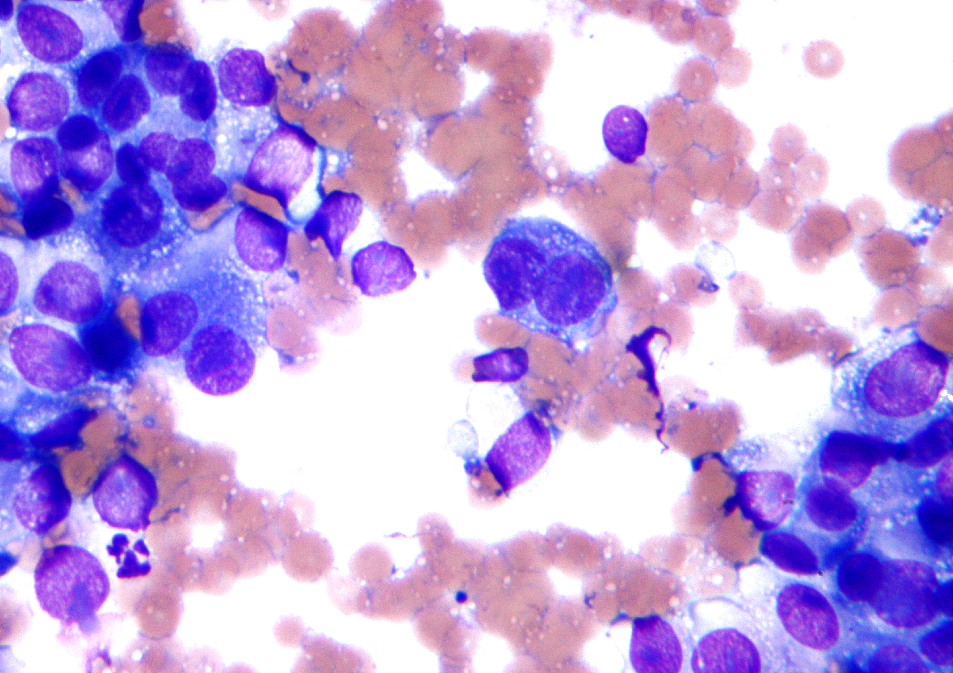
Description: Micrograph of malignant melanoma. Cytology specimen. Field stain. The micrograph shows features commonly seen in melanoma: Large (>2x the size of a RBC), dyscohesive cells. Epithelioid binucleated single cells, "bug-eyed monster cells". Cells with: Large nucleoli. Abundant granular cytoplasm. Features associated with melanoma but not seen: Pigment. Pseudoinclusions. Singular spindle cells.
Title: Melanoma - cytology field stain
Credit: Own work
Author: Nephron
Usage Terms: Creative Commons Attribution-Share Alike 3.0
License: CC BY-SA 3.0
License Link: https://creativecommons.org/licenses/by-sa/3.0
Attribution Required?: Yes
Image usage
The following page links to this image:

All content from Kiddle encyclopedia articles (including the article images and facts) can be freely used under Attribution-ShareAlike license, unless stated otherwise.