
Image: Panoramic radiograph of historic dental implants
Size of this preview: 800 × 536 pixels. Other resolutions: 320 × 214 pixels | 1,350 × 904 pixels.
Original image (1,350 × 904 pixels, file size: 98 KB, MIME type: image/jpeg)
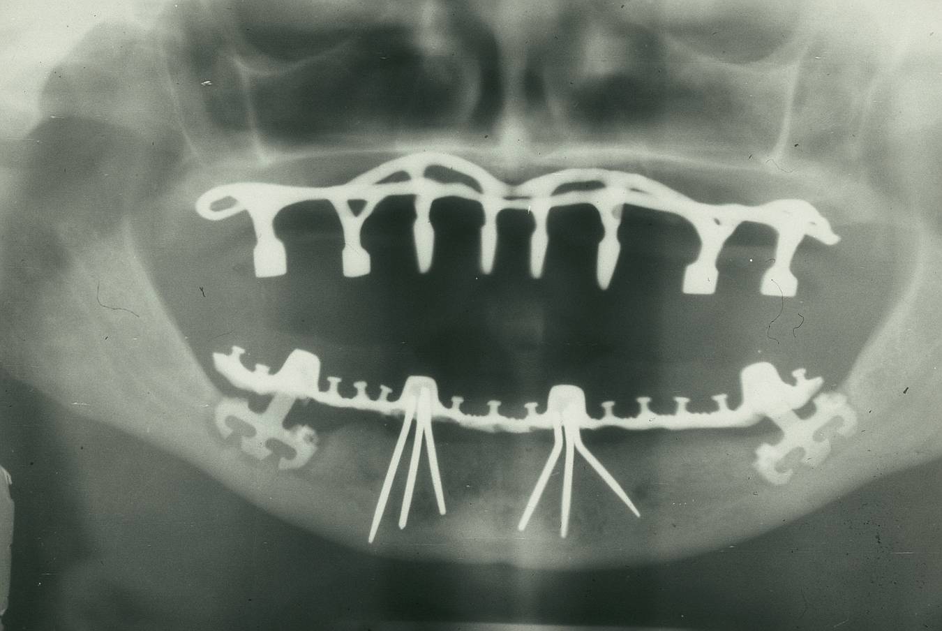
Description: The x-ray controlling from 1976/77 shows an subperiosteal implant (according Cherchéve) in the maxilla. Two implant tripods (according Pruin) in the lower canine region and two stabilized blade Implants (according Heinrich) in the molar region.
Title: Panoramic radiograph of historic dental implants
Credit: Own work
Author: Dentistxxx
Usage Terms: Creative Commons Attribution-Share Alike 3.0
License: CC BY-SA 3.0
License Link: http://creativecommons.org/licenses/by-sa/3.0
Attribution Required?: Yes
Image usage
The following page links to this image:

All content from Kiddle encyclopedia articles (including the article images and facts) can be freely used under Attribution-ShareAlike license, unless stated otherwise.