
Image: Sickle cell 01
No higher resolution available.
Sickle_cell_01.jpg (375 × 600 pixels, file size: 100 KB, MIME type: image/jpeg)
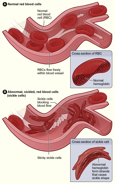
Description: Figure A shows normal red blood cells flowing freely in a blood vessel. The inset image shows a cross-section of a normal red blood cell with normal hemoglobin. Figure B shows abnormal, sickled red blood cells blocking blood flow in a blood vessel. The inset image shows a cross-section of a sickle cell with abnormal (sickle) hemoglobin forming abnormal strands.
Title: Sickle cell 01
Credit: http://www.nhlbi.nih.gov/health/health-topics/topics/sca/
Author: The National Heart, Lung, and Blood Institute (NHLBI)
Usage Terms: Public domain
License: Public domain
Attribution Required?: No
Image usage
The following page links to this image:

All content from Kiddle encyclopedia articles (including the article images and facts) can be freely used under Attribution-ShareAlike license, unless stated otherwise.