
Image: X-ray of pacemaker with right atrial and ventricular lead
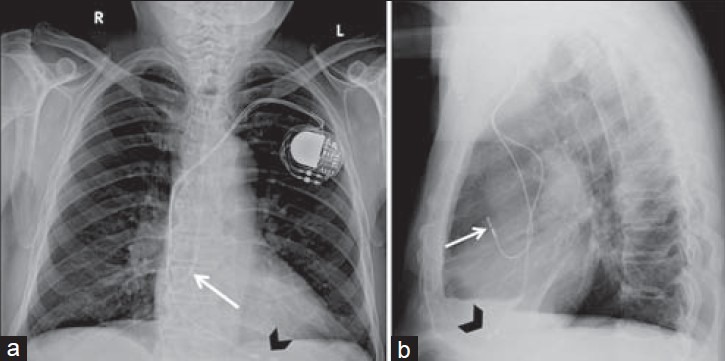
Description: Dual-chamber pacemaker. 89-year-old male with a dual-chamber pacemaker placed via the left subclavian vein with the generator in the subcutaneous left infraclavicular fossa. Chest X-ray (a) PA view and (b) LAT view show the right atrial lead is inferiorly directed and takes a “J loop” curve with tip pacing the right atrial appendage (white arrow). Right ventricle lead directed inferiorly through the right atrium and tricuspid valve, then courses anteriorly and inferiorly with the tip at the right ventricular apex (black arrowhead). In single-chamber pacemakers, only the right atrial or ventricular lead is present.
Title: X-ray of pacemaker with right atrial and ventricular lead
Credit: (2014). "Radiography of Cardiac Conduction Devices: A Pictorial Review of Pacemakers and Implantable Cardioverter Defibrillators". Journal of Clinical Imaging Science 4 (1): 74. DOI:10.4103/2156-7514.148269. ISSN 2156-7514.
Author: Stephanie C Torres-Ayala, Guido Santacana-Laffitte, and José Maldonado
Usage Terms: Creative Commons Attribution 4.0
License: CC BY 4.0
License Link: https://creativecommons.org/licenses/by/4.0
Attribution Required?: Yes
Image usage
The following 2 pages link to this image:
