
Scanning electron microscope facts for kids


The scanning electron microscope (SEM) is a type of electron microscope that uses a focused beam of high-energy electrons in producing a variety of signals at the surface of a solid specimen . The signals produced by the interacting electrons contain useful information such as the shape, atomic structure and conductivity.
When an electron hits the surface, it may be reflected (backscattered), absorbed, or conducted away. Electrons that are absorbed can cause the atom that they hit to become unstable, forcing it to give off another electron (a secondary electron), or to give off light in order to stabilize. Different detectors, for the different types of reactions, may be fitted to an electron microscope, depending on what is being looked for.
The magnification that can be achieved in a scanning electron microscope depends on how narrow the beam of electrons that strikes the surface can be, and can reach 1 nanometer, about the size of 3 to 5 atoms. The control of the beam is achieved using magnetic fields, with other magnetic fields being used to shape the beam, and to move it across the sample. The range of magnification may range from 30x to as high as 500,000x.
The electron beam is affected by air and water molecules, so the sample must be placed in a vacuum. The sample must also be conductive to allow the electrons not reflected or absorbed to be conducted away. This gives some limits on the type of sample that can be used. In the case of samples from plants or animals, it often means that they must first be coated in gold before they can be placed in the microscope. This is done in order to preserve the sample and keep it from changing or decaying throughout the scanning process.
The scanning electron microscope is made up of several components:
1. The electron gun is located at the very top or bottom of an SEM and is used to fire a beam of electrons at the object that we are looking at under SEM.
2. The lenses are used to focus electron beams so that we can get clear and detailed images of the examined object. They are not made of glass, but they are made of magnets that change the path of electrons created from the electron gun.
3. The sample chamber is the area where the specimen of interest is examined under SEM. As the technique is very sensitive, the chamber must be very sturdy and have no vibrations happen. This is to allow for the specimen to be kept very still in order for the SEM to produce good images.
4. The detector is the "eyes" of the microscope. It is able to produce the most detailed images of a specimen's surface. Many different detectors can be used depending upon the information wanted from the sample used.
5. The vacuum chamber is used to keep the electron beam from constant interference from air. The air particles are bad for electron beams in that the electrons would be knocked out of the air and onto the specimen, which will produce bad distorted surfaces of the specimen.
Images for kids
-

Schottky-emitter electron source
-
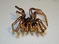
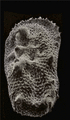
A spider sputter-coated in gold, having been prepared for viewing with an SEM
-

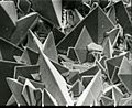
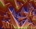
Low-temperature SEM magnification series for a snow crystal. The crystals are captured, stored, and sputter-coated with platinum at cryogenic temperatures for imaging.
-
Comparison of SEM techniques: Top: backscattered electron analysis – composition Bottom: secondary electron analysis – topography
-
Color cathodoluminescence overlay on SEM image of an InGaN polycrystal. The blue and green channels represent real colors, the red channel corresponds to UV emission.
-
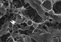
Surface of a kidney stone
-
The same after re-processing of the color from the estimated topography
-
SEM image of a diagenetically altered discoaster
-
The same image after similar colorization
-
SEM image of Cobaea scandens pollen
-

The same after semi-automatic coloring. Arbitrary colors help identifying the various elements of the structure
-

Colored SEM image of Tradescantia pollen and stamens
-
Colored SEM image of native gold and arsenopyrite crystal intergrowth
-
DDC-SEM of calcified particle in cardiac tissue - Signal 1 : SE
-
Signal 2 : BSE
-

Colorized image obtained from the two previous. Density-dependent color scanning electron micrograph SEM (DDC-SEM) of cardiovascular calcification, showing in orange a calcium phosphate spherical particle (denser material) and, in green, the extracellular matrix (less dense material)
-
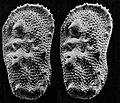
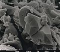
An SEM stereo pair of microfossils of less than 1 mm in size (Ostracoda) produced by tilting along the longitudinal axis.
-
From this pair of SEM images, the third dimension has been reconstructed by photogrammetry (using MountainsSEM software, see next image) ; then a series of 3D representations with different angles have been made and assembled into a GIF file to produce this animation.
-
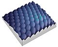
3D surface reconstruction of a (Ra = 3 µm) roughness calibration sample (as used to calibrate profilometers), from 2 scanning electron microscope images tilted by 15° (top left). The calculation of the 3D model (bottom right) takes about 1.5 second and the error on the Ra roughness value calculated is less than 0.5%.
-

SEM image of a house fly compound eye surface at 450× magnification.
-

Detail of the previous image.
-
SEM 3D reconstruction from the previous using shape from shading algorithms.
-

Same as the previous, but with lighting homogenized before applying the shape from shading algorithms
-

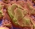
Colored SEM image of soybean cyst nematode and egg. The artificial coloring makes the image easier for non-specialists to view and understand the structures and surfaces revealed in micrographs.
-
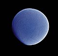
Compound eye of Antarctic krill Euphausia superba. Arthropod eyes are a common subject in SEM micrographs due to the depth of focus that an SEM image can capture. Colored picture.
-

Ommatidia of Antarctic krill eye, a higher magnification of the krill's eye. SEMs cover a range from light microscopy up to the magnifications available with a TEM. Colored picture.
-
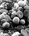
SEM image of normal circulating human blood. This is an older and noisy micrograph of a common subject for SEM micrographs: red blood cells.
-
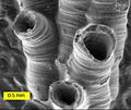
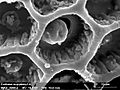
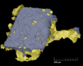
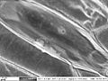
SEM image of a hederelloid from the Devonian of Michigan (largest tube diameter is 0.75 mm). The SEM is used extensively for capturing detailed images of micro and macro fossils.
-
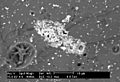
Backscattered electron (BSE) image of an antimony-rich region in a fragment of ancient glass. Museums use SEMs for studying valuable artifacts in a nondestructive manner.
-
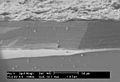
SEM image of the corrosion layer on the surface of an ancient glass fragment; note the laminar structure of the corrosion layer.
-
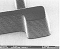
SEM image of a photoresist layer used in semiconductor manufacturing taken on a field emission SEM. These SEMs are important in the semiconductor industry for their high-resolution capabilities.
-
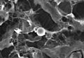
SEM image of the surface of a kidney stone showing tetragonal crystals of Weddellite (calcium oxalate dihydrate) emerging from the amorphous central part of the stone. Horizontal length of the picture represents 0.5 mm of the figured original.
-
Two images of the same depth hoar snow crystal, viewed through a light microscope (left) and as an SEM image (right). Note how the SEM image allows for clear perception of the fine structure details which are hard to fully make out in the light microscope image.
-
Epidermal cells from the inner surface of an onion flake. Beneath the shagreen-like cell walls one can see nuclei and small organelles floating in the cytoplasm. This BSE-image of a lanthanoid-stained sample was taken without prior fixation, nor dehydration, nor sputtering.


.gif)


See also
 In Spanish: Microscopio electrónico de barrido para niños
